INTRODUCTION TO THE STANLEY BRAIN COLLECTION
E. Fuller Torrey and Maree Webster, Stanley Medical
Research Institute, Bethesda, Maryland
The Stanley Brain Collection was established in 1994 to
collect postmortem brain tissue from individuals with schizophrenia, bipolar
disorder, major depression, and normal controls. At this time, the
collection includes 430 specimens, approximately evenly divided among these four
diagnoses. The tissue is collected by selected collaborating medical
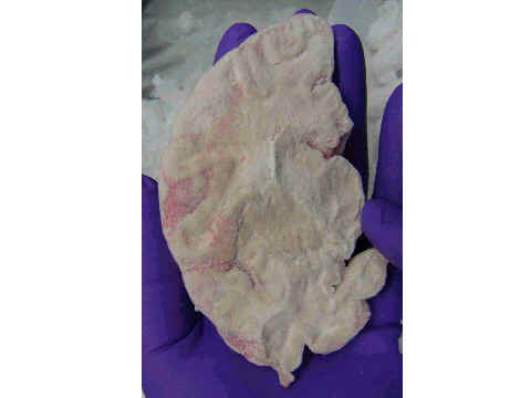
examiners, with permission of the family, and processed in a uniform
manner. One half of the brain is frozen, and the other half is fixed in
formalin. Diagnoses are established by review of medical records and, when
needed, interviews with family members.
A collection of 60 brains, 16 matched from each of the four
diagnostic groups has been made available to researchers. Called the
Neuropathology Consortium, over 100,000 blocks and sections of the Consortium
have been sent to 120 researchers around the world without charge. We are
now considering what kind of a collection to create to be used primarily for
microarray analysis. Would it be better to have a smaller closely matched
case-control series, e.g., 12 in each group? Or a less-well-matched but much
larger series, e.g., 40 in each group? And what should the relative
priorities be for matching for such things as age, sex, race, pH, mode of death,
interval from death to refrigeration, interval from date to freezing of tissue,
and freezer storage time?
How should priorities for tissue allocation be
established? Since microarrays require tissue blocks for the extraction of
RNA, there is a very limited amount of time in some structures of great interest
such as the hippocampus, amydala, and nucleus accumbens.
Slides:
Maree Webster
 |
E. Fuller Torrey
 |
 |
|
 |
 |
 |
 |
 |
 |
 |